RNA microscope maps tumor in 3D
How cells organize themselves in a tissue is crucial to understanding how they work together – and how childhood cancer develops. Benedetta Artegiani and Delilah Hendriks are working on a new method to map tumor tissue in 3D. Not with a traditional microscope, but by analyzing the spatial information in RNA.
To understand how cells in a tissue communicate with each other, knowledge of their spatial organization is essential. 3D information can also show “islands” within a tissue of cells with different properties. This can provide insight, for example, into why a tumor only partially responds to treatment.
Research group leaders at the Princess Máxima Center, Benedetta Artegiani and Delilah Hendriks, are therefore working on a technique that uses DNA and RNA information to examine biological information of tumors directly in their 3D environment.
Thin slices
To view a tumor in 3D, researchers can use a microscope with fluorescent markers that make certain cells light up. With new technologies, for example, called spatial transcriptomics, they can look at genetic information at a deeper level. But in both methods, the tissue must first be cut into thin slices. So the tissue does not remain intact, and 3D resolution is lost – and with it, some biological information.
Spatial information without cutting
Together with an international consortium, Artegiani and Hendriks are working on a way to apply spatial transcriptomics directly to human tumor tissue in 3D. ‘We analyze the unique molecular fingerprint of each cancer cell in the tumor. This provides insight into the activity and location of each cell. But we do this with a crucial difference: without cutting up the tissue to extract the DNA and RNA from each cell,’ explains Delilah Hendriks, co-leader of the Artegiani & Hendriks group. ‘Instead, we use networks of molecular information that allow us to determine the position of each cell in our 3D model.’
Future: improving diagnosis and treatment
A research consortium led by the KTH Royal Institute of Technology in Sweden is carrying out the project, called Voluminex. The team recently received a prestigious Pathfinder Open grant, part of the European Innovation Council’s Horizon Europe program.
Artegiani and Hendriks plan to apply the technique in their research on the brain tumor organoids they developed in their group. ‘By using the spatial information in DNA, we hope to find a cost-effective way to map tumors and organoids three-dimensionally,’ said Benedetta Artegiani, co-leader of the Artegiani & Hendriks group. ‘This method can not only help study the interaction between tumor and healthy cells, but also be applied in the clinic for tumor analysis of each child. Fast, affordable 3D tumor snapshots can improve diagnosis, and possibly even lead to new treatments.’
The Voluminex consortium consists of five partners: KTH Royal Institute of Technology, the Princess Máxima Center, Karolinska Institutet, Single Technologies AB and Sorbonne University. The project received a grant of €3 million.

Utrecht Science Park Summer Café: Farewell to Managing Director Jan Henk van der Velden
On Monday, 6 July, the Utrecht Science Park Summer Café took place at Grand Café Living on Utrecht Science Park. This edition was dedicated to the farewell of Jan Henk van der Velden, Managing Director of the Utrecht Science Park Foundation, after nine years in which he played a key role in the development and positioning of the ecosystem.
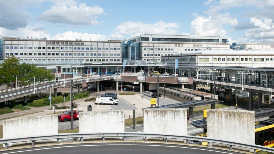
UMC Utrecht is to get a new main entrance and a green campus park
The north side of UMC Utrecht is set to undergo a major redevelopment in the coming years. Utrecht City Council and the teaching hospital have presented plans for a new main entrance, a central car park and a green reception area designed to improve connections between the hospitals at Utrecht Science Park. The plans have now been submitted to the city council.

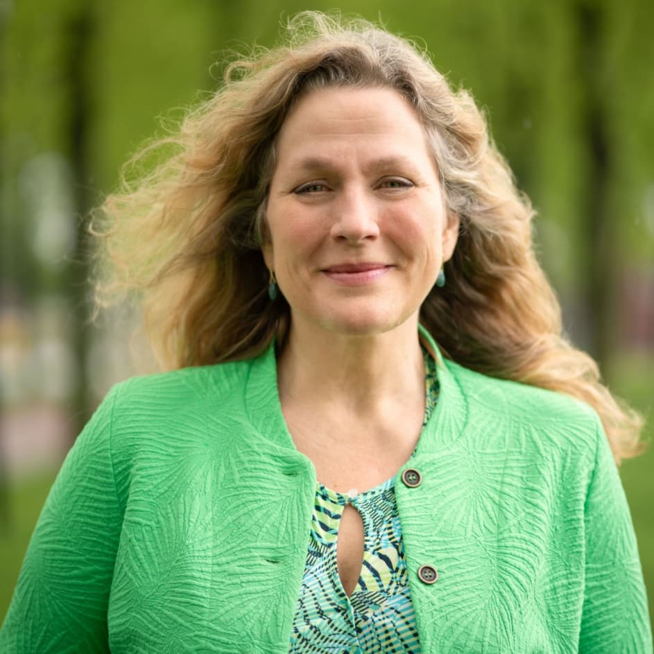
Petra Baarendse appointed new Managing Director of Stichting Utrecht Science Park as of 1 September
Utrecht, 2 July 2026 – As of 1 September 2026, Petra Baarendse will take office as the new Managing Director of Stichting Utrecht Science Park. She succeeds Jan Henk van der Velden, who has stepped down and will take on a new role as Managing Director of Stichting Nationaal Park Utrechtse Heuvelrug.
Opening Utrecht Science Week with keynote by Juliette Legler on the hidden influence of environmental chemicals
On Friday, September 25, the annual science festival ‘Utrecht Science Week’ kicks off once again at Utrecht Science Park. We are pleased to announce that this year, Prof. Dr. Ir. Juliette Legler will deliver the Utrecht Science Lecture. Juliette Legler is Professor of Toxicology at the Faculty of Veterinary Medicine at Utrecht University and leads the ‘One Health Toxicology’ group at the Institute for Risk Assessment Sciences (IRAS).