Measuring brain blood flow during brain surgery
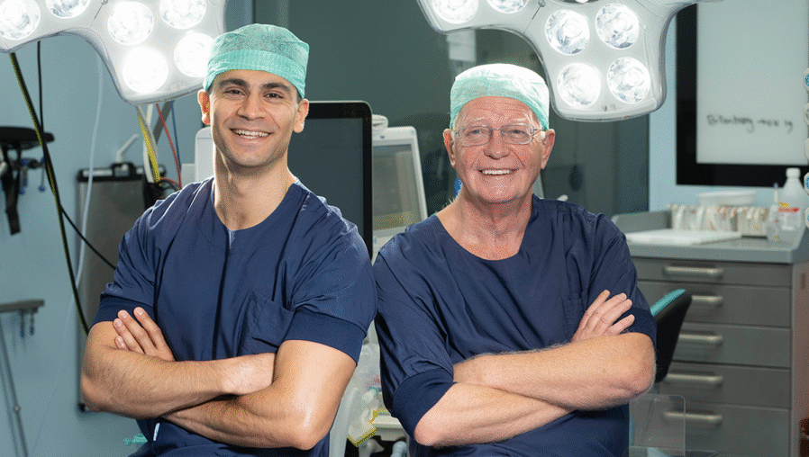
Neurosurgeons at UMC Utrecht use a new ultrasound technique to monitor brain blood flow live during surgery, enabling earlier detection and prevention of stroke risks.
Neurosurgeons at UMC Utrecht can, for the first time, monitor brain tissue blood flow live during brain surgery thanks to a new technique. Together with researchers from TU Eindhoven, they have developed a method to detect—and potentially prevent—the risk of a stroke during surgery. The technique could also be useful for other operations, such as kidney transplants.
During surgeries on brain vessels, for example when treating an aneurysm or creating a bypass, there is a risk that blood flow to the brain tissue temporarily stops. This can lead to a stroke. The risk starts at around 8 percent in aneurysm surgeries and can rise to nearly 50 percent in complex cases. For brain tumors like gliomas, the risk ranges between 12.5 and 44 percent.
Pilot with ten patients
Until now, surgeons had no tool to directly observe a stroke during surgery. Thanks to a special form of ultrasound imaging—Ultrafast Power Doppler Imaging (UPDI)—this has changed. The technique visualizes tiny blood vessels, measures blood volume in the brain tissue, and shows live what happens during the procedure. Surgeons can, for example, immediately see if the brain tissue receives enough blood when a blood vessel is temporarily clamped. The technique is fast, safe, and easy to apply during surgery.
Researchers at UMC Utrecht tested the technique on ten patients undergoing brain vessel surgery. During the operation, they continuously measured changes in blood flow using the new ultrasound setup. In some cases, they clearly saw the tissue receiving less or temporarily more blood right after releasing a vessel clamp. “We observed effects that otherwise only become visible afterward—when it’s already too late—such as on an MRI scan. Now we can measure this during surgery,” says neurosurgeon Dr. Dara Niknejad, who led the research.
The technique could potentially be used in many other surgeries, Dara adds. “It’s useful for any operation where continuous tissue blood flow is important. We haven’t tried it yet, but think, for example, of kidney transplants: the surgeon can immediately see if the organ is fully perfused after transplantation. UPDI can also improve organ assessment before transplantation.”
Adaptation of the imaging technique
Professor of biomedical signal processing Massimo Mischi from TU Eindhoven adds: “The technology and expertise for using UPDI in this setting were developed in our Biomedical Diagnostics lab. Dr. Yizhou Huang succeeded in using and processing high-frequency, ultrafast ultrasound signals from multiple angles. This provided surgeons with high-resolution images of brain blood flow.”
The precise, high-resolution measurements of blood flow in the brain enable early detection of reduced perfusion, says Massimo. “This allows surgeons to take timely precautions during surgery and prevent serious complications such as stroke.”
Fewer complications, better control
Thousands of patients in the Netherlands undergo brain surgery annually, for example for aneurysms, brain tumors, or vascular malformations. Such surgeries sometimes require temporary closure of a brain blood vessel, increasing the risk of stroke. With this new technique, researchers hope to better manage this risk. “We want to prevent complications as much as possible. By monitoring blood flow in real-time, we can intervene more quickly if something goes wrong,” Dara explains.
The pilot is a first step. In further research, the team wants to measure how well the technique can predict whether a patient actually suffers a stroke. They also want to explore how to further integrate the technique into the operating room. Dara: “Our ultimate goal is to make brain surgery safer, with fewer complications and better outcomes for patients.”

Registration for Utrecht Science Week 2026 is now open: the knowledge festival at Utrecht Science Park
From Friday 25 September to Sunday 4 October, the sixth edition of Utrecht Science Week will take place. Utrecht Science Park will open its doors to everyone interested in (applied) science and innovation in the fields of life sciences, health, and sustainability.
The first programme elements have now been announced and registration is open. Many more events and activities will be added to the programme in the coming weeks.
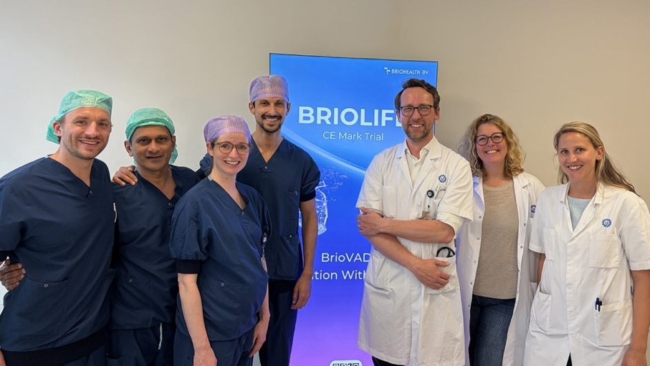
New artificial heart implanted for the first time in Europe
Earlier this month at UMC Utrecht, a patient with advanced heart failure became the first person in Europe to receive a BrioVAD ventricular assist device. The new system is being evaluated in a European study and may offer advantages over the existing ventricular assist device. For UMC Utrecht, the implantation marks a new milestone in a long history of innovation in the field of mechanical circulatory support.
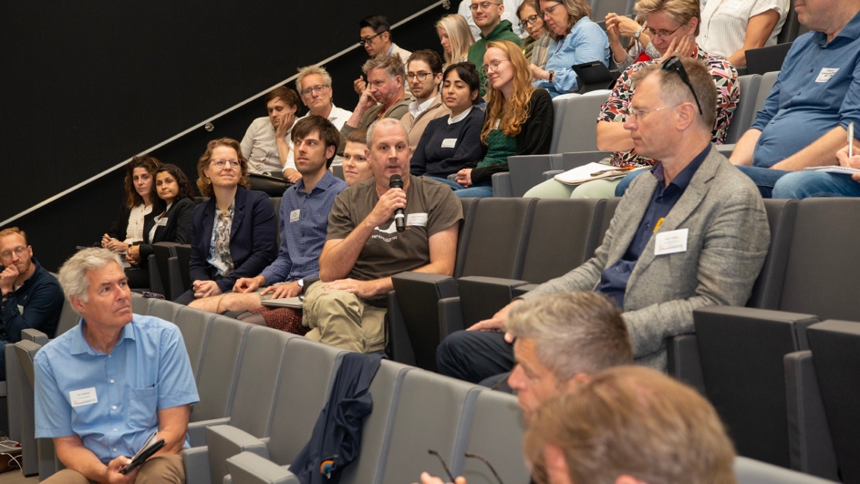
Successful first edition of Utrecht Life Sciences TechConnect
On 4 June 2026, researchers, technology experts, facility staff and innovation partners from across the Utrecht Life Sciences community gathered at Utrecht Science Park for the first edition of Utrecht Life Sciences TechConnect. The new annual event was created to strengthen connections between researchers and technology facilities, showcase advanced research technologies, and stimulate collaboration across disciplines.
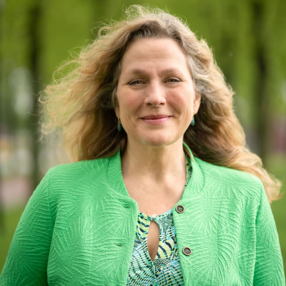
Opening Utrecht Science Week with keynote by Juliette Legler on the hidden influence of environmental chemicals
On Friday, September 25, the annual science festival ‘Utrecht Science Week’ kicks off once again at Utrecht Science Park. We are pleased to announce that this year, Prof. Dr. Ir. Juliette Legler will deliver the Utrecht Science Lecture. Juliette Legler is Professor of Toxicology at the Faculty of Veterinary Medicine at Utrecht University and leads the ‘One Health Toxicology’ group at the Institute for Risk Assessment Sciences (IRAS).